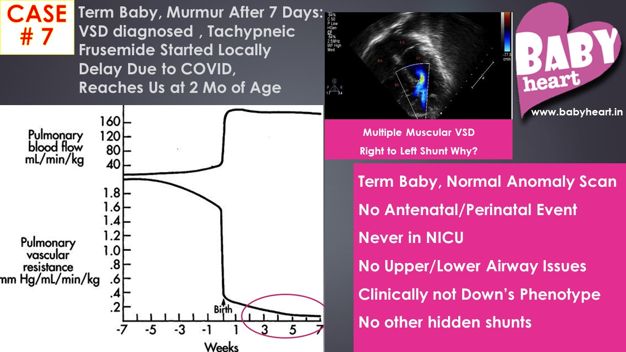
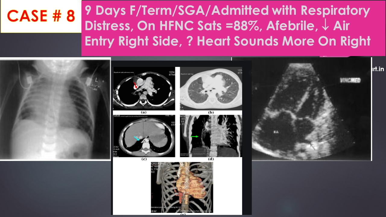
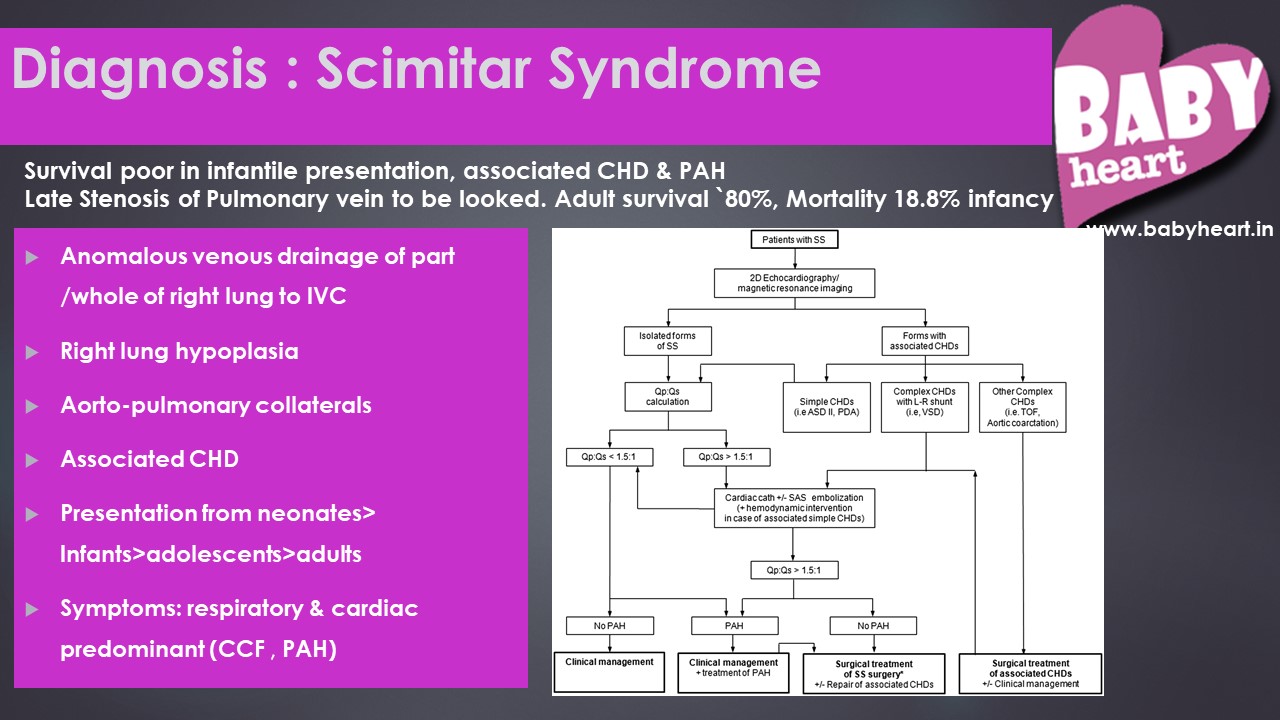
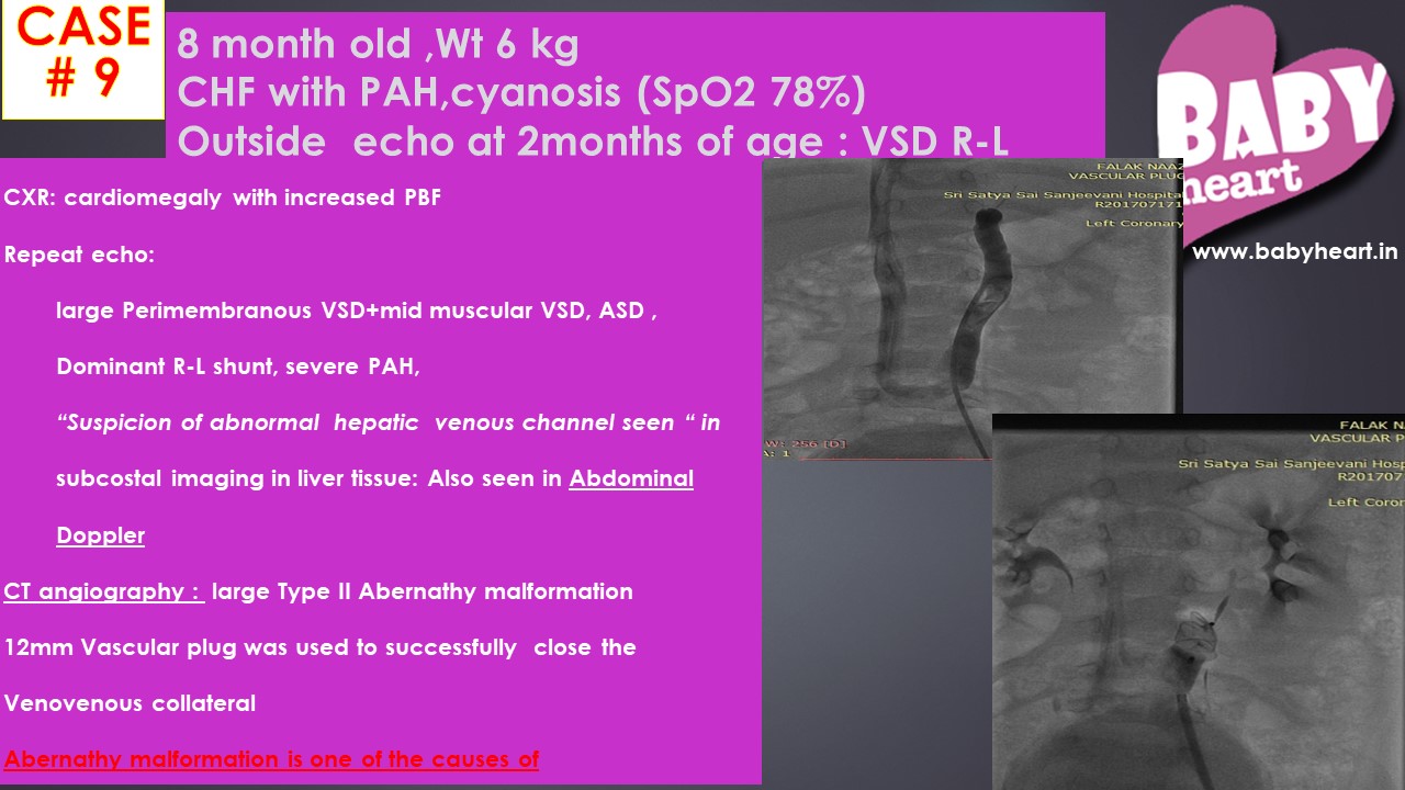
Hole in the Heart: Quick Recap for Parents
By Justin
Every year 8-12 children per 1000 live deliveries are born with heart defects. These are also called Congenital Heart Defect (CHD) which simply means that the heart developed problems since when it was being formed inside the womb. The commonest problems are holes in the heart. Although sound innocuous, this short phrase in my experience created a lot of apprehension and fear among the family and also health care providers. This article is our attempt to put the facts straight.
The Normal Heart:

Click on the image to see a larger version
The heart is a pumping organ and has got four chambers. The two upper chambers are called the Right Atrium and Left Atrium (RA and LA) and the two lower chambers are called the Right Ventricle and Left Ventricle (RV and LV). The upper chambers are separated by a wall called inter-atrial septum and the lower chambers are separated by another wall called inter-ventricular septum. From the RV, one artery or pipe takes impure /deoxygenated blood to lungs for oxygenation. This is called the Pulmonary Artery (PA) and from the LV another artery called Aorta carries pure/oxygen-rich blood to the whole body.
Continue to common holes in the heart –>
What to do if your child has a hole in the heart?
By Maitri
There is no simple and/or single answer to this. Your best guide is the pediatric cardiologist. As I have told that all of us are born with small ASD and PDA. Usually they close spontaneously by 1st week of life. Premature babies often have large PDAs and they often close after medical therapy with oral Ibuprofen or Indomethacin. Rarely some preemies need surgery. The baby’s neonatologist will start these.
Regarding ASD, small defects upto 3-5mm, often close spontaneously. However, larger defects like more than 5-8mm size do not close. They need closure by 3-4 years of age i.e. preschool age either by operation or non-operatively by a button called DEVICE. This is a nonsurgical technique, done under general anesthesia and takes about 40-45 minutes. The child is observed for 24 hours post procedure and then discharged. The advantage is this that there is no scar and unless someone does an Echo or Chest X-ray, it is impossible to identify that the child had an ASD. The usual hospital stay is 36 hours.
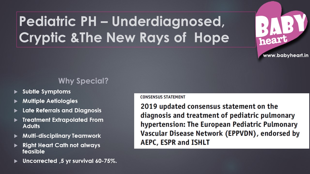
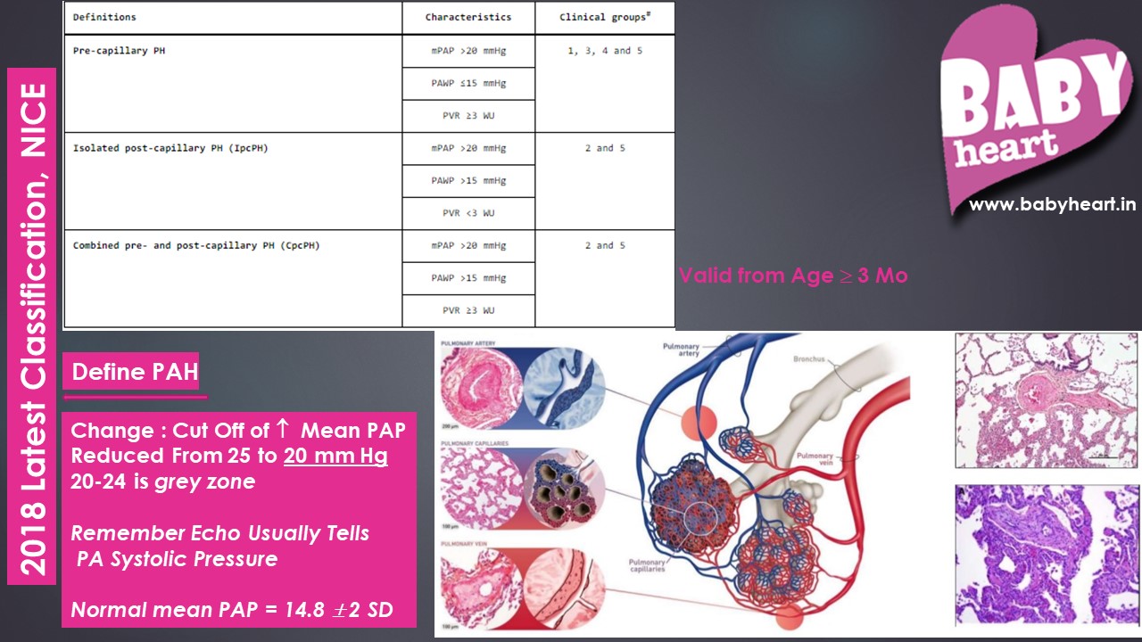
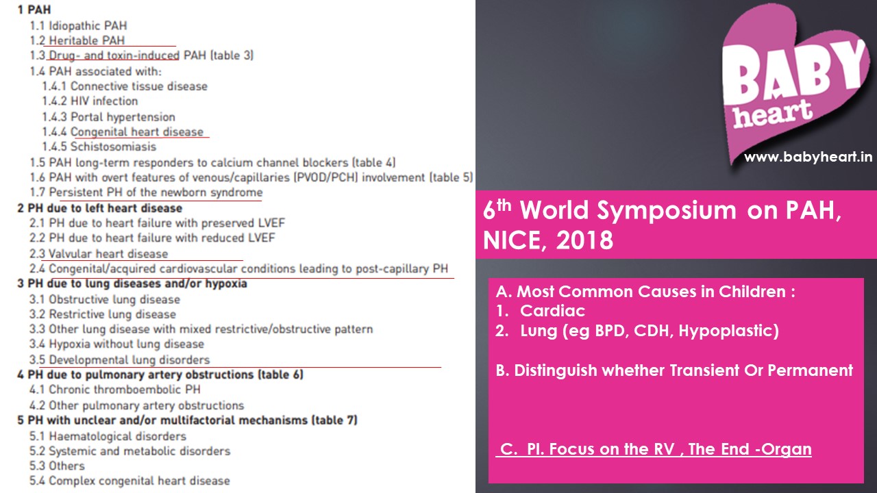
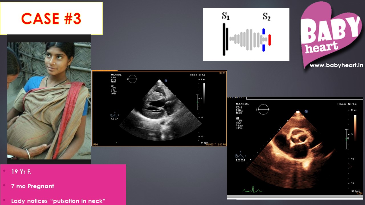
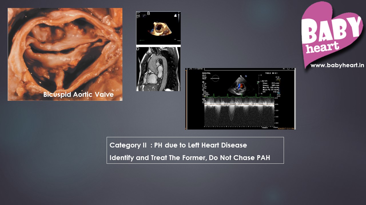
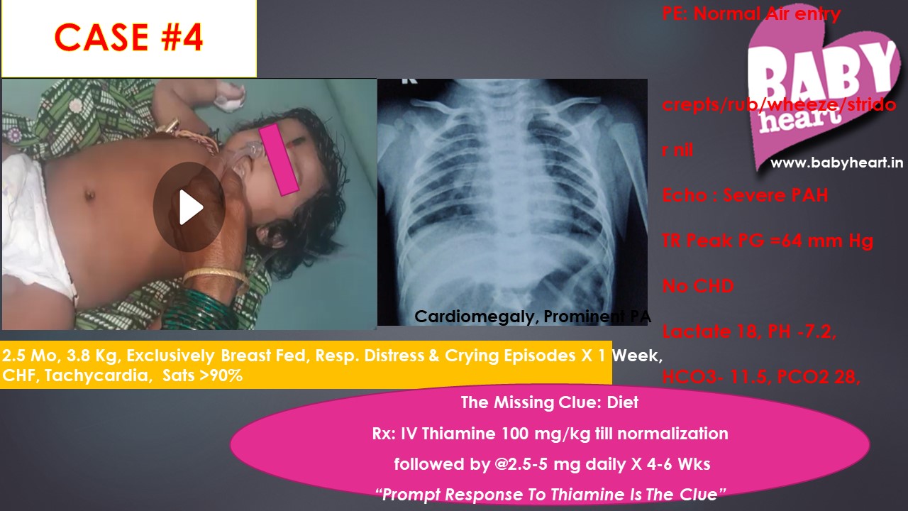
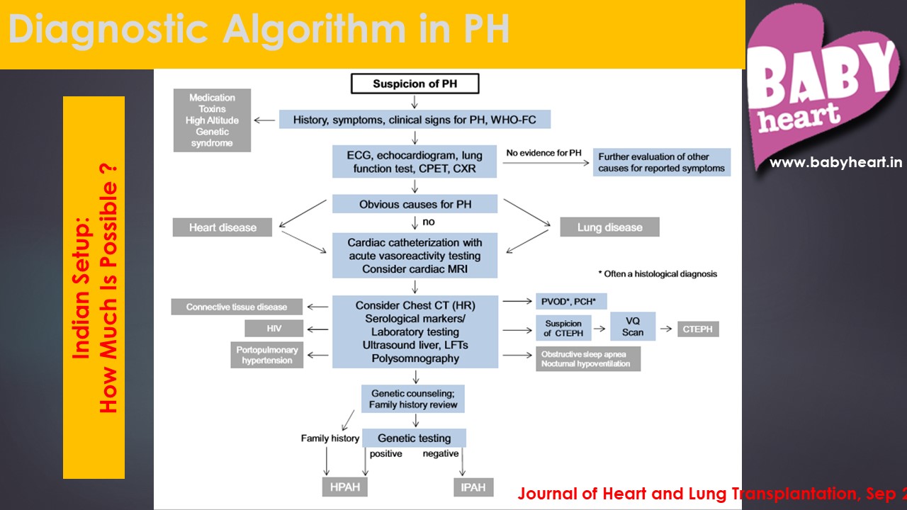
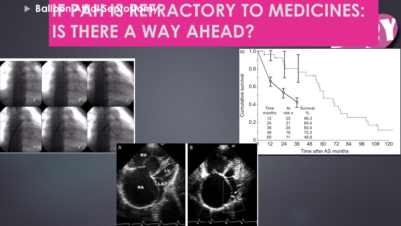
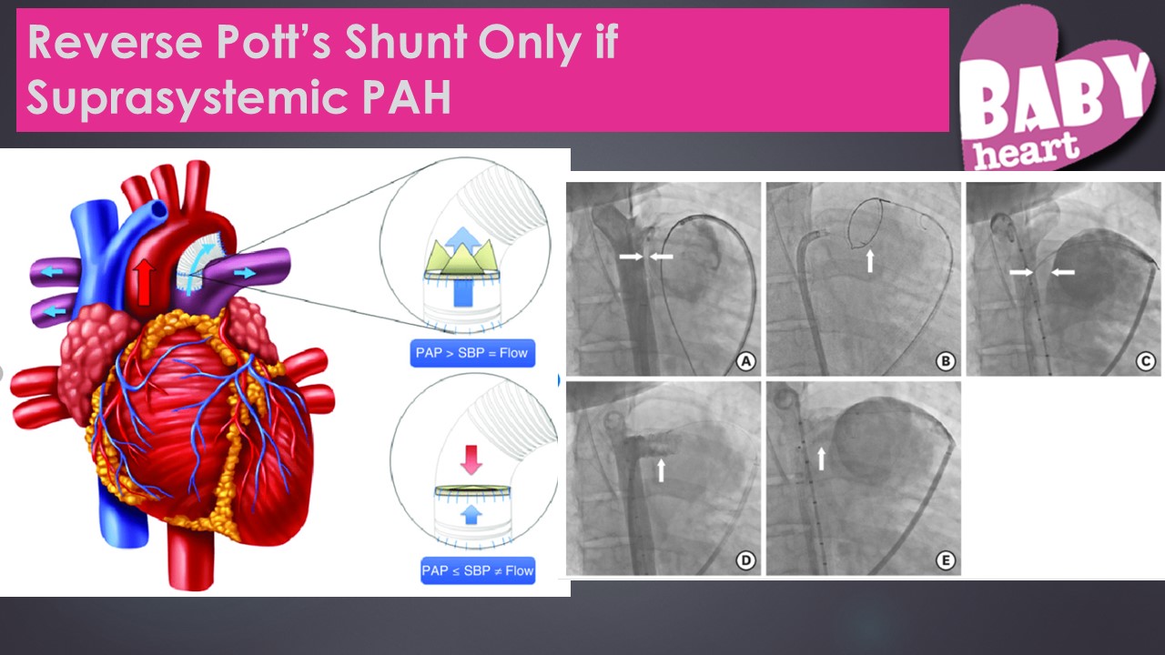
Pulmonary Hypertension in Children – Recent updates & Indian scenario
By Maitri
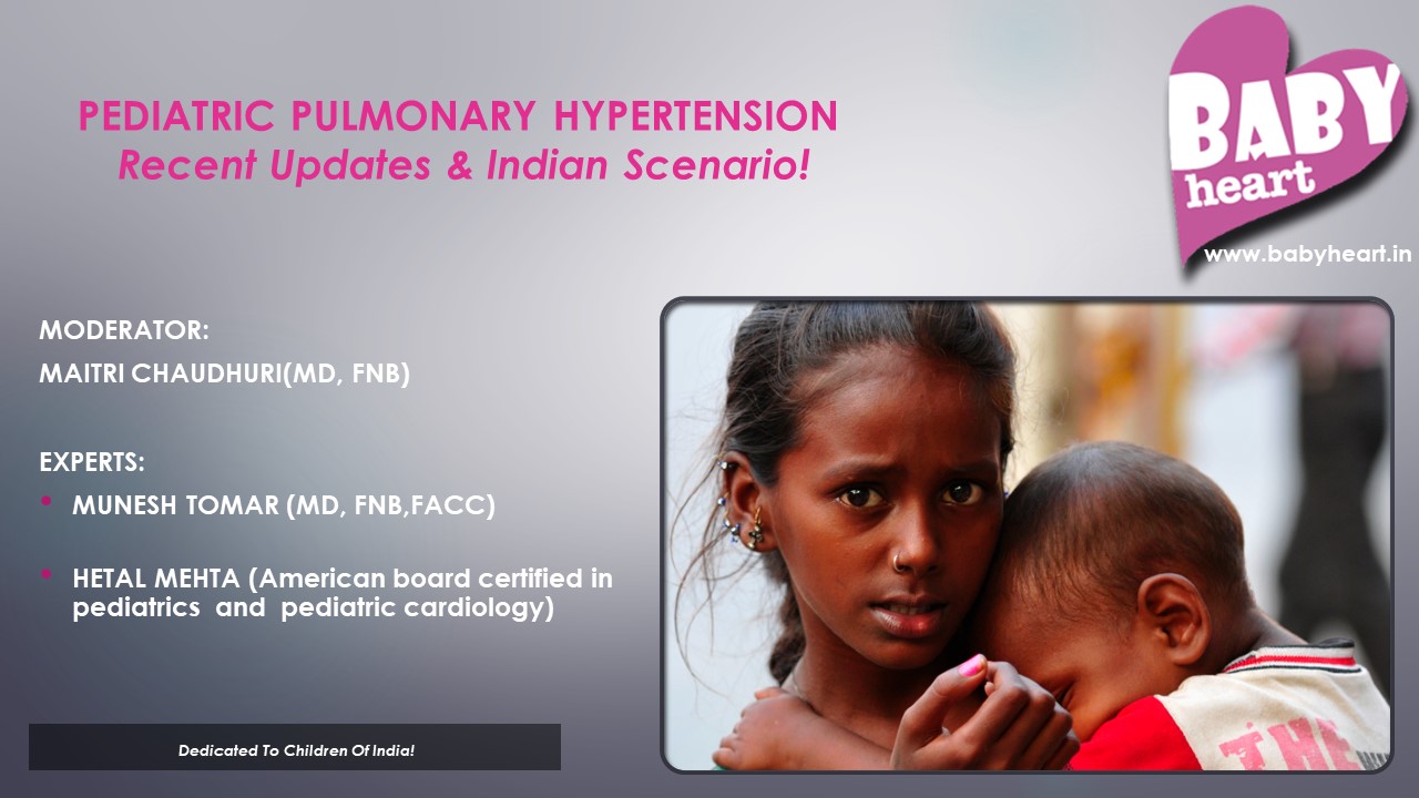
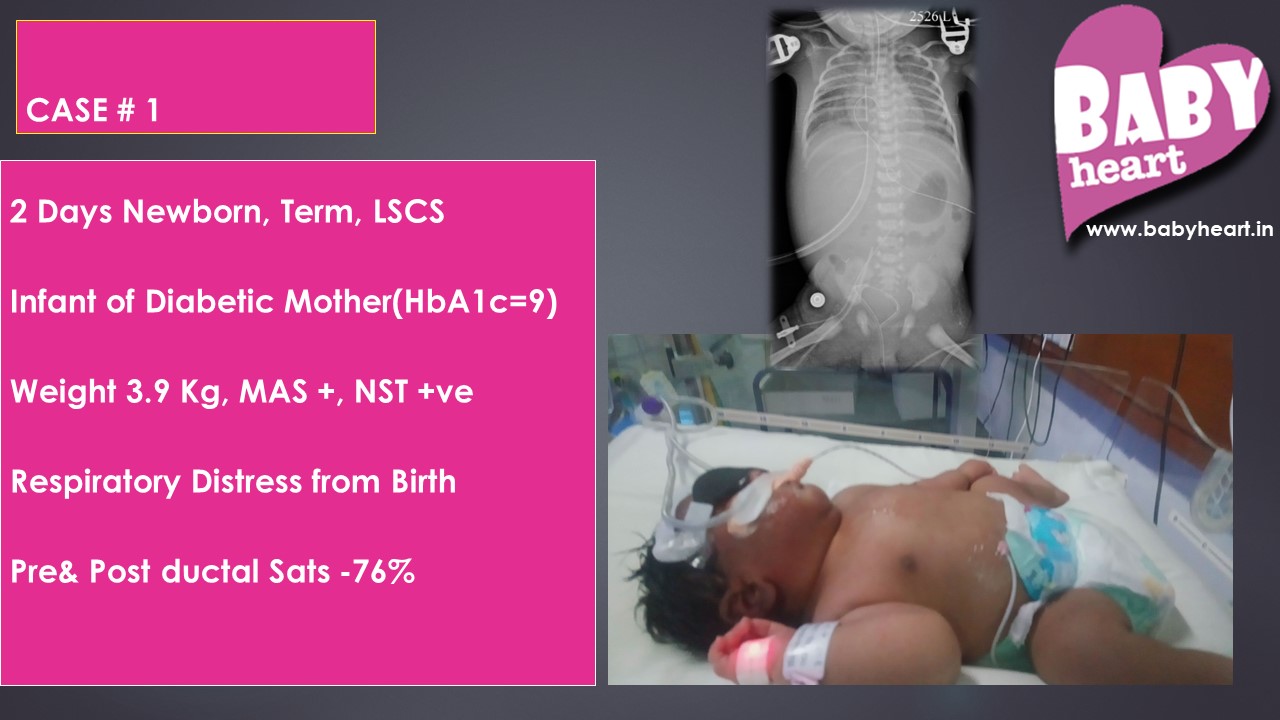
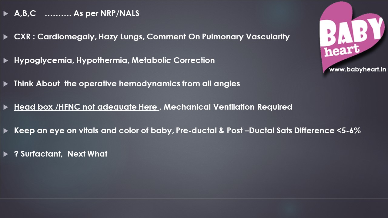
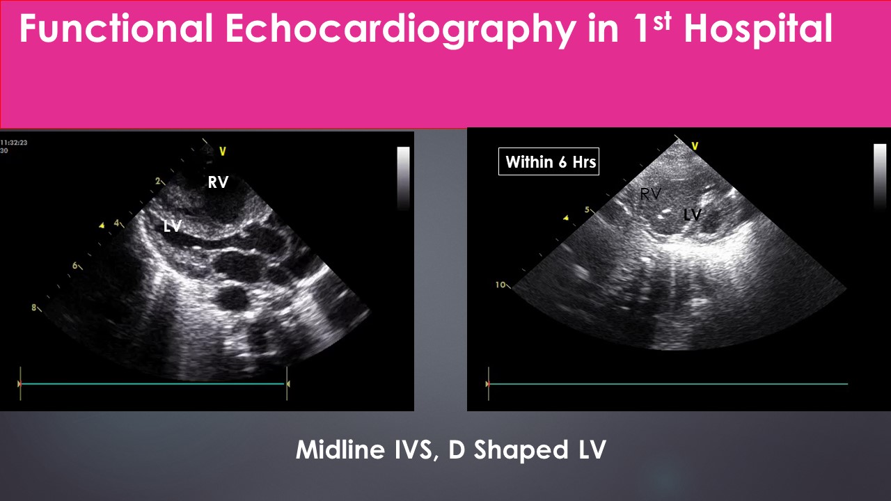
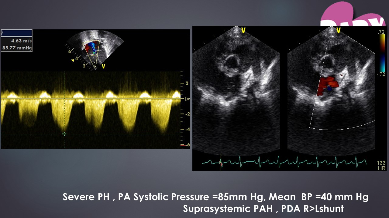
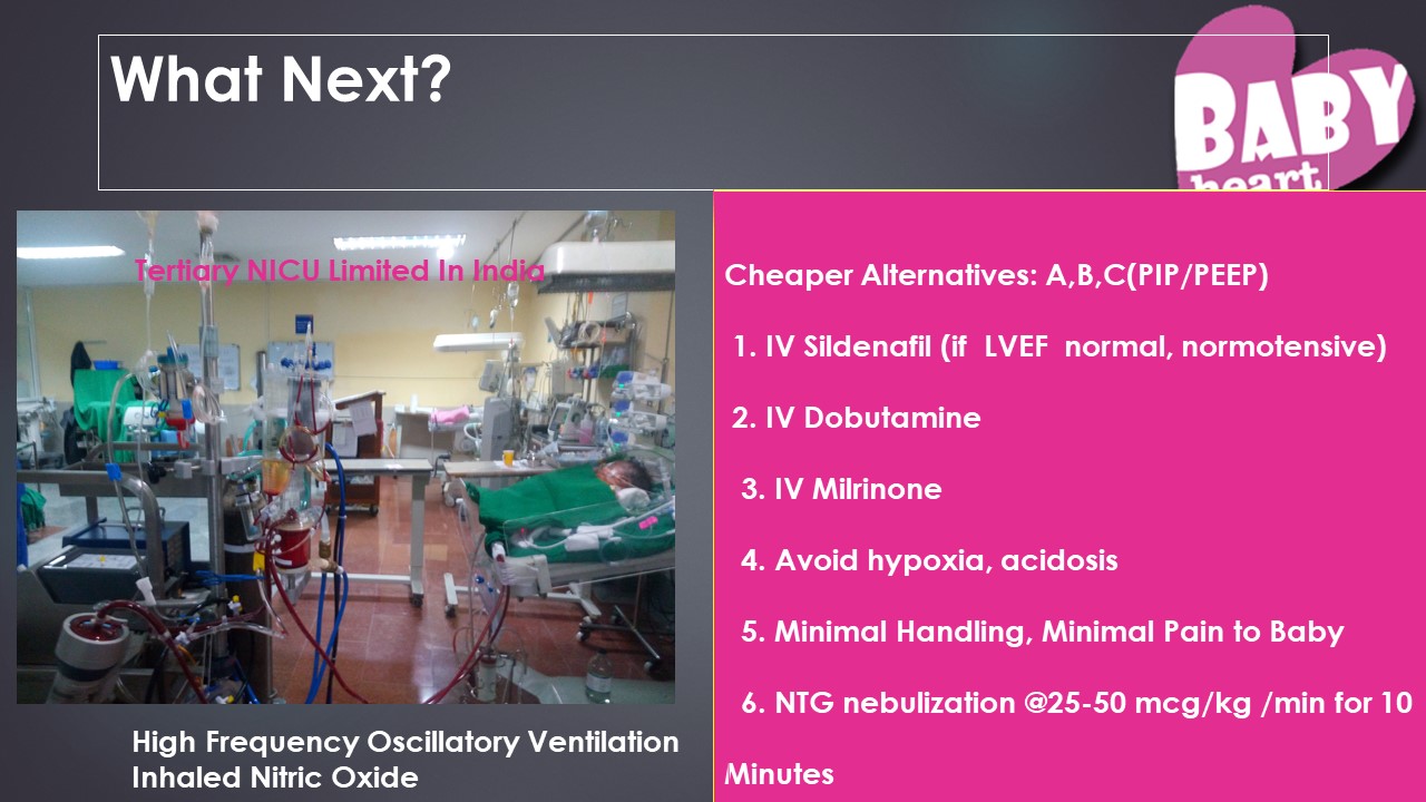
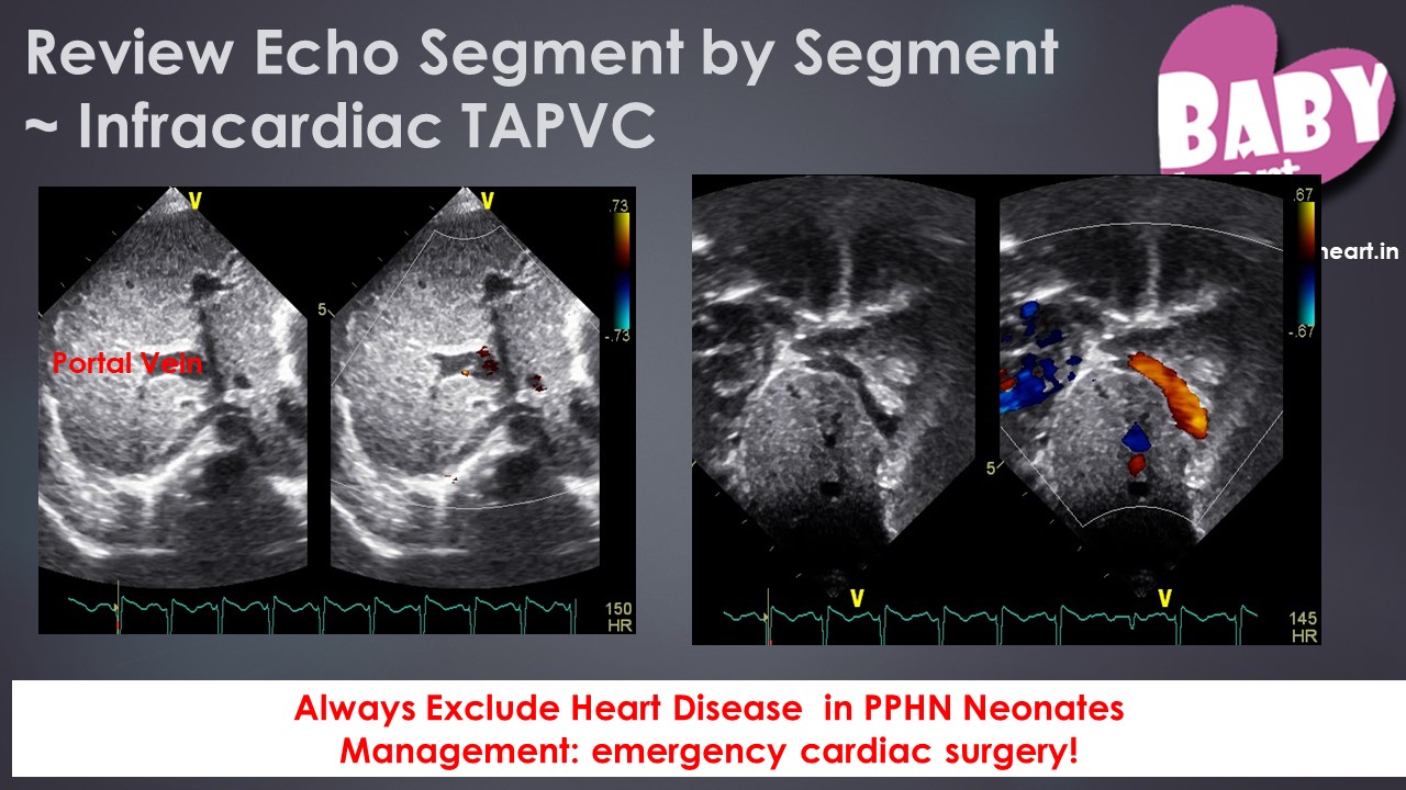
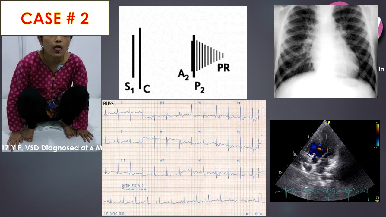
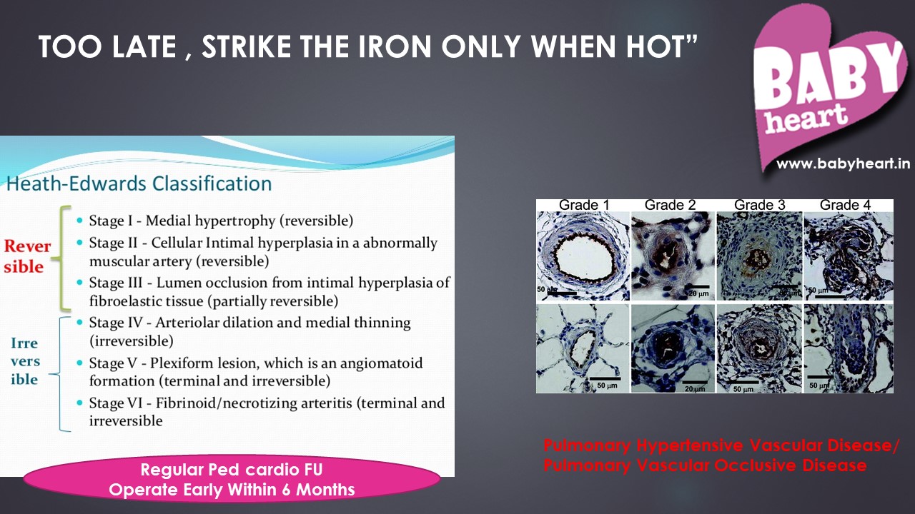
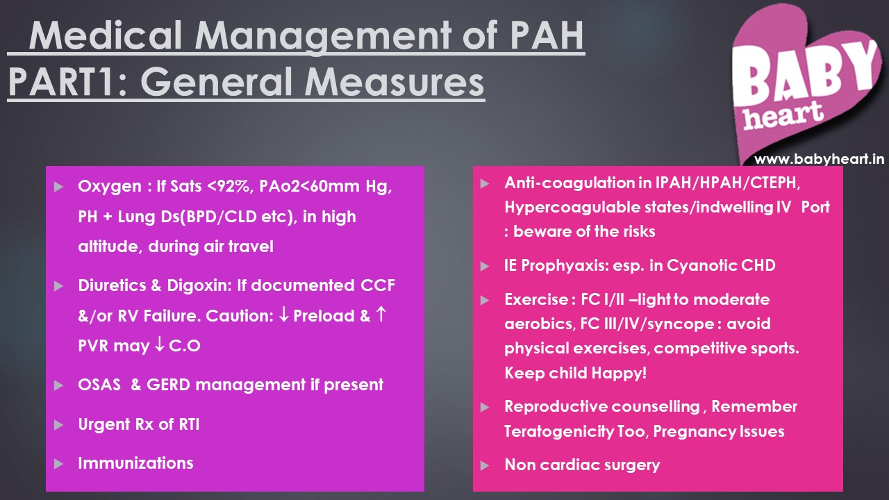
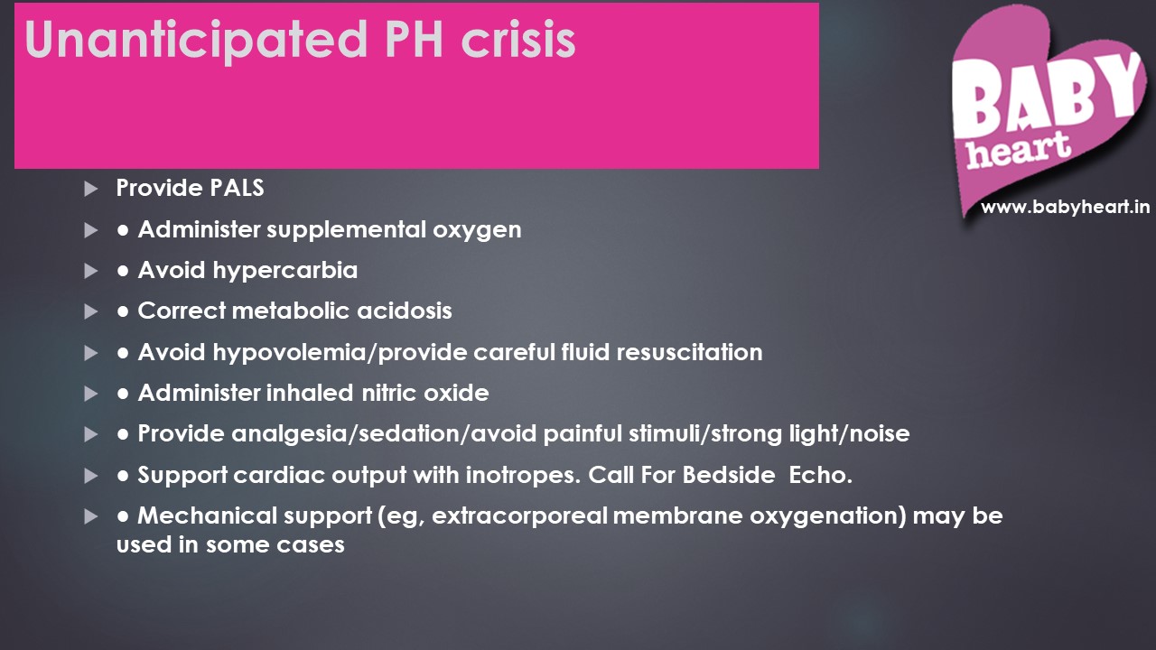
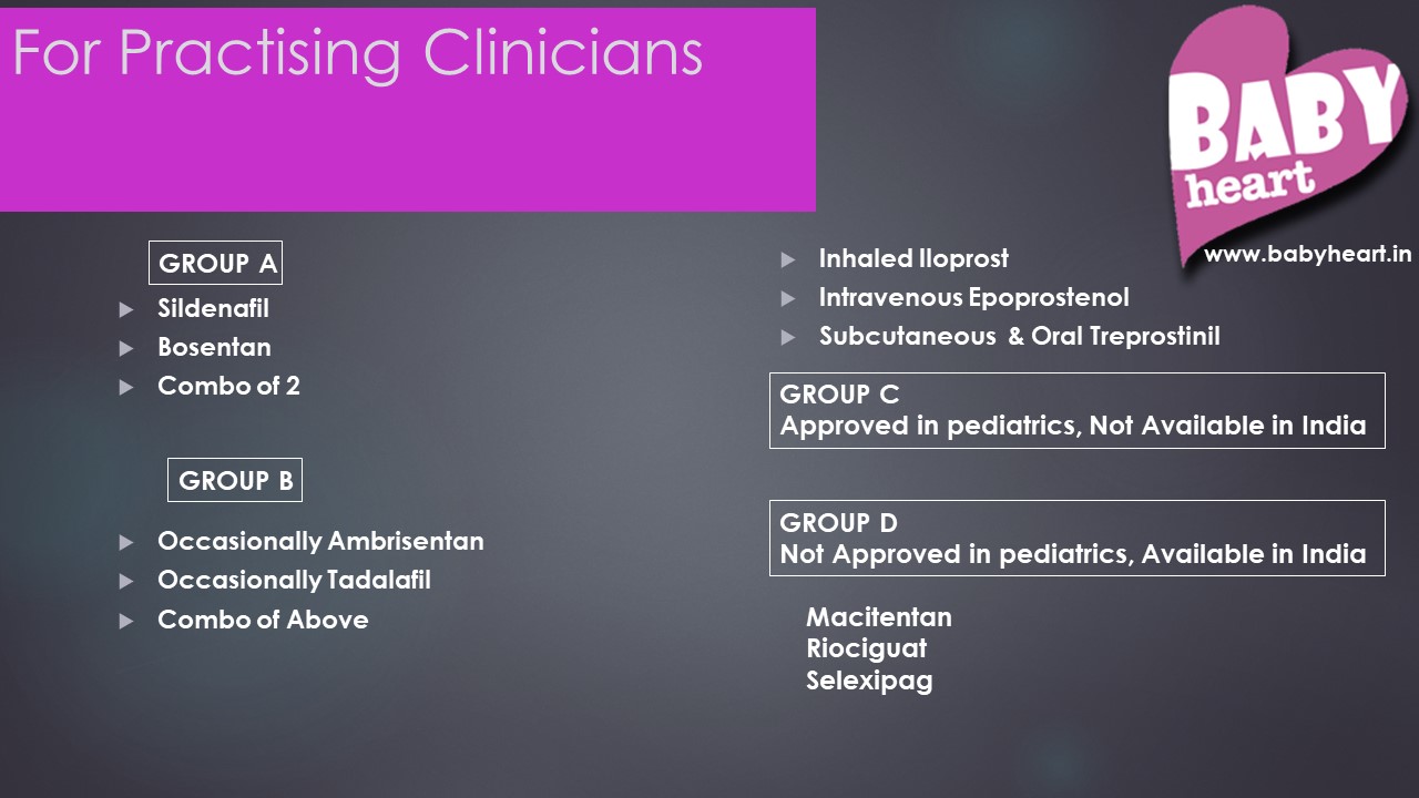
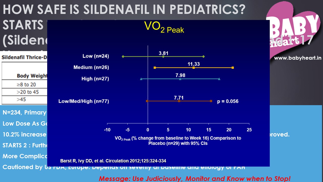
Pulmonary hypertension in Children: Recent updates and multiple Indian scenarios are discussed in the webinar by Dr Maitri Chadhuri. Please click on an image to start slideshow. If you would like to download the whole PowerPoint presentation with comments and notes, please see below the gallery for a download link


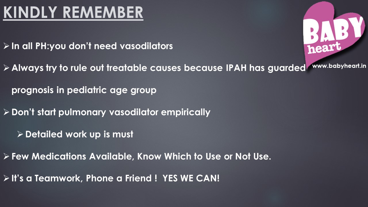

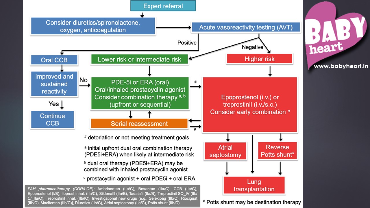
Dr. Maitri Chaudhuri
Click here to download PowerPoint presentation (PPT) with comments and notes or click here to download as a printable PDF
Pediatric and Fetal Echo Z-Score Calculators
By Justin
The free Pediatric and Foetal echo Z score calculators allow you to generate z-scores and reference values (normal values) for Pediatric and Foetal echo using data compiled from medical literature and publications
Click to select the Calculator you want to use
Aortic Root Z-Scores
Calculate BSA-adjusted z-scores of the aortic annulus and sinuses of Valsalva using data published by Boston Children’s Hospital. (Updated 11/2008: added percentiles)
Cardiac Valve Z-Scores
Calculate BSA-adjusted z-scores of the mitral valve, tricuspid valve, aortic valve, and pulmonary valve using data published by Cincinnati Children’s Hospital. This data is also used to create dynamic z-score tables.
M-Mode Z-Scores
Calculate BSA-adjusted z-scores for common M-Mode measurements (LVEDD, LVESD, IVS, LVPW, LA) using data published by Johannes Gutenberg University, Germany.
TAPSE
Reference values for right ventricular systolic function, using tricuspid annular plane systolic excursion (TAPSE).
Z-Scores of Cardiac Structures: Detroit Data
Calculate BSA-adjusted z-scores of 21 common pediatric echo measurements (m-mode, mitral valve, aortic valve, pulmonary arteries, etc.) using data published by Detroit Children’s Hospital. (Updated 11/2008: added percentiles)
Z-Scores of Cardiac Structures: Wessex Data
Calculate BSA-adjusted z-scores of 15 cardiac structures (mitral valve, aortic valve, pulmonary arteries, etc.) using data published by the Wessex Cardiothoracic Unit, Southampton UK.
Thanks to Dan Dyar and Parameter(z).com for this wonderful tool. All the source codes used for Z score calculations in babyheart are adapted from it. Z score calculator by Dan Dyar is licensed under a Creative Commons Attribution 3.0 License and is based on a work at parameterz.com
Kawasaki Disease comprehensive presentation
By Maitri
Kawasaki disease is an Idiopathic multisystem disease characterized by vasculitis of small & medium blood vessels, including coronary arteries. This video includes everything from history of the disease to treatment plans and options.